
Managing a Single Central Crown
Introduction
Replacing a single central crown or veneer is tricky. About as tricky as shade matching gets, in my opinion of course. We have all heard the phase centrals should be twins, laterals siblings and canines cousins. But how exactly can we get the centrals to be identical, especially when often the underlying stump is so dark. In this post I will discuss a recent case where this was managed.


History
This gentleman was referred to me by a colleague regarding his current central crown which was placed many years ago. The tooth was root filled and crowned following trauma and has been asymptomatic ever since. He has attended as he would like the crown replacing, improving the aesthetics of his smile. He does not want a smile makeover per say, but would like his two front teeth to match.
Consent
With any case such as this consent plays a large part. When removing old work , even with radiographs we are never certain what is present underneath. Looking at this periapical radiograph although there is no associated infection and the bone levels are good there is a potential that the post has fractured. This may have resulted in the root being fractured and subsequently the tooth may be unrestorable, we need our patients to understand this prior to embarking on treatment.
Treatment
On the day of treatment, the existing crown was removed. It is in this moment when your heart sinks as you see caries and a stump as dark as the shades of night. Fortunately the post was rock solid. An attempt was made to remove the post however I did not want to cause more damage with its removal and therefore I elected to leave the post.

To Mask or Not to Mask?
A common question I get asked is should I mask the dark stump? This depends mainly on the restorative plan. Zirconia is historically opaque, however as the science develops more translucent zirconias exist. My take on it is, especially in this case where there is LOADs of space, if we mask the stump it gives the lab more options. Moreover as teeth are naturally translucent, even with staining, you can often notice a difference if the opacity is vastly different. In this case the caries was removed and the margin pushed apically to the gum line. After this everything was sandblasted and bonded with scotchbond (3M). First a pink opaquer (3M) was used, followed by white opaquer (DMG) then finally a A3 body dentine shade (Venus) placed to mimic a dentine layer underneath. The prep was refined using a red grit torpedo bur (Schottlander).

Scanning & Lab Instructions
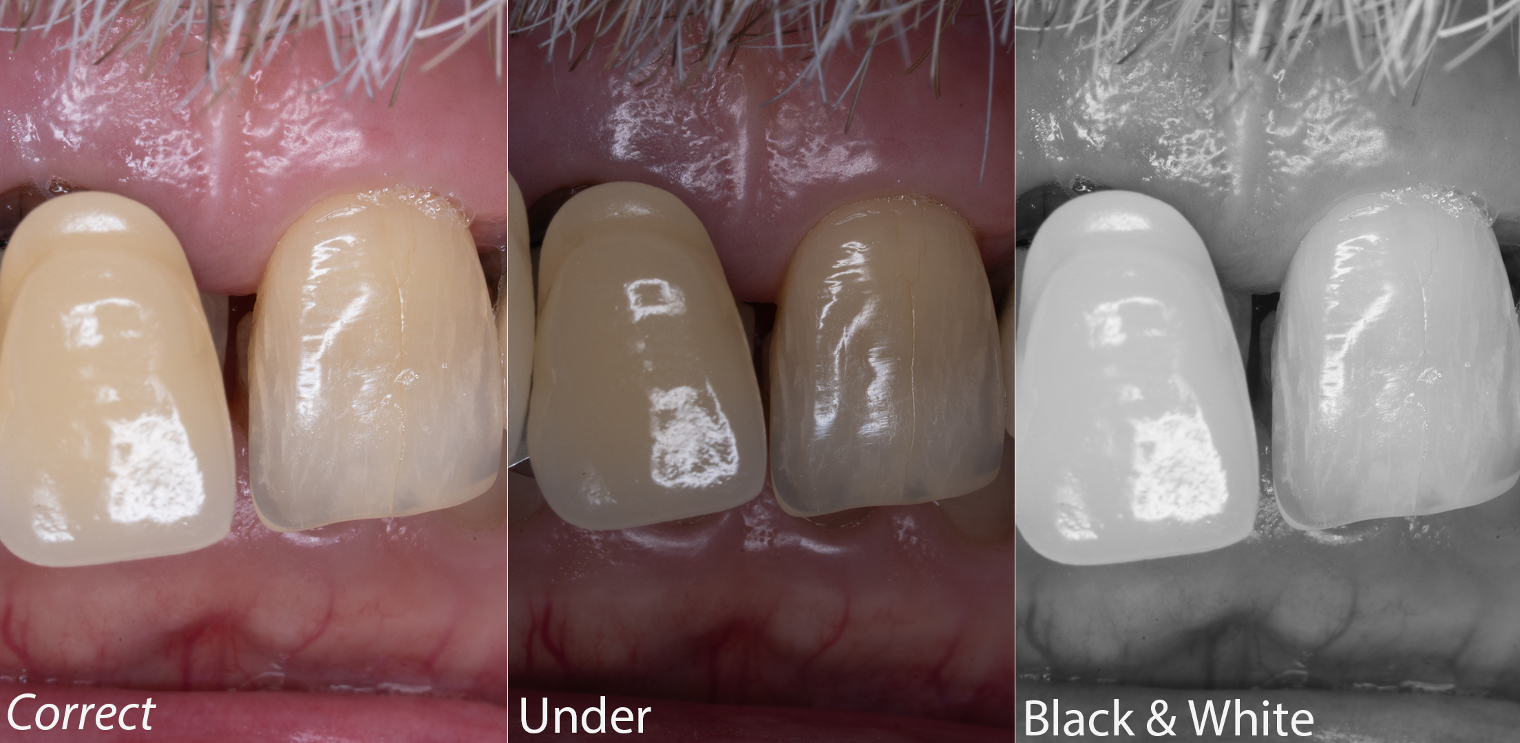
After preparation, a two cord technique was used with a 00 cord initially used followed by placement of a 0 cord (Ultrapack). The 0 cord was removed prior to intra-oral scanning with a trios 3 IOS. Records were sent to the lab, annotated on an iPad. When tackling a case such as this, I like to send a variety of photographs in different lights mainly:
Correct exposure - Shows what the tooth is like in normal light
Under exposure - Shows lots of details in the tooth
Black and White - Shows value well

Try In
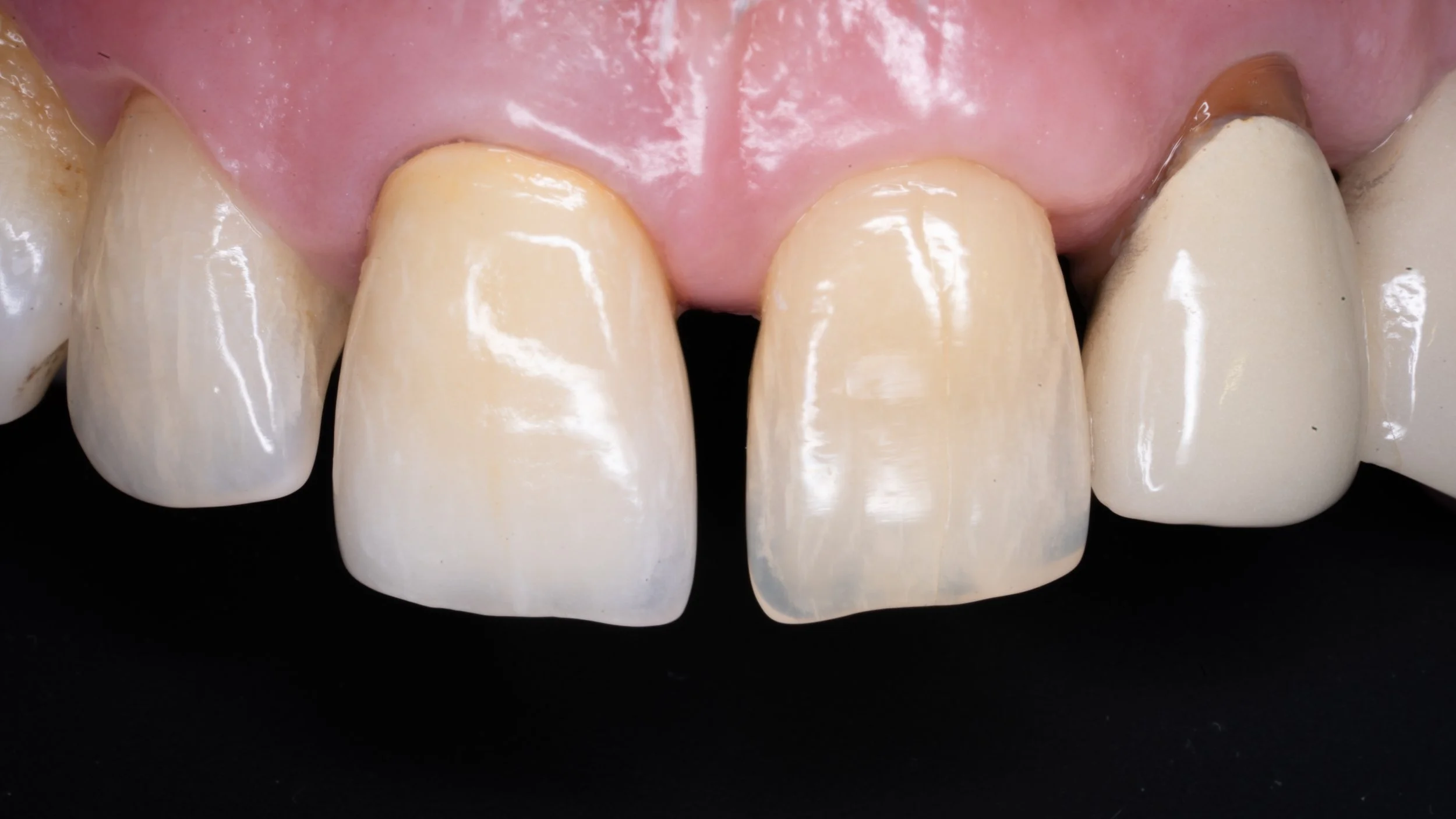
The scans, lab work and images were sent to the incredible Byrnes Lab (Click Here) and the first crown was produced. With single centrals I never book a “fit appointment” especially with the first crown as chances are some adjustments are required. This manages the patients expectations prior to embarking on the treatment. The crown arrived back and I loved the shape and general characterisation although I felt the value was too high, especially in the incisal third. You might also notice the margin on show, this was due to my temporary being over extended in this area. As I had sounded to bone at the start of the treatment I was confident that the gingivae would bounce back once the temporary was adjusted.
Fit
The crown was sent back with the instructions to lower the value in the incisal third. The crown returned for a second time and was much improved. The prep was sandblasted. As the crown was zirconia, it was cleaned internally with ivoclean and luted in with relyx unicem. Being super picky there is a discrepancy in the halo between the two incisors and it is ever so slightly longer than its partner however I feel this is a lovely result for a very nice patient.
